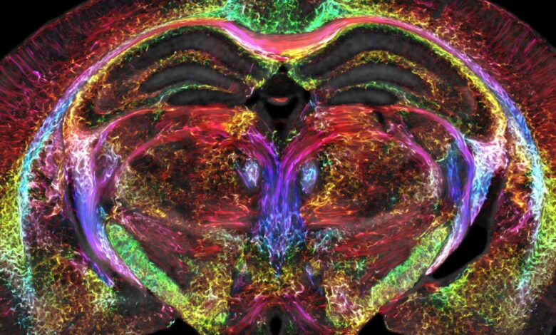
A super-powerful MRI merged with light-sheet microscopy allows researchers to create a high-definition wiring diagram of the entire brain in mice. Credit: Duke Centre for In Vivo Microscopy
HealthTechnology USASharper MRIs: Brain Scans Are Looking Clearer Than Ever
Following decades of efforts to improve the resolution of Magnetic Resonance Imaging (MRI), a team of researchers from diverse American universities has created the sharpest images yet of a mouse brain, leading to a better understanding of the human brain.
“It is something that is truly enabling. We can start looking at neurodegenerative diseases in an entirely different way,” explains G. Allan Johnson, Ph.D., the lead author of the new paper and the Charles E. Putman University Distinguished Professor of radiology, physics and biomedical engineering at Duke.
Scientists from the University of Tennessee Health Science Center, the University of Pennsylvania, the University of Pittsburgh, and Indiana University, under the leadership of Duke Center for In Vivo Microscopy, created dramatically crisper images made of voxels 64 million times smaller than a clinical MRI voxel. To achieve this feat, the team used a powerful 9.4 Tesla magnet instead of a 1.5 to 3 Tesla magnet, a unique set of gradient coils 100 times stronger than clinical MRIs, and a high-performance computer equivalent to 800 laptops. It is now possible to visualize the connectivity of the entire brain at a record-breaking solution, and better understand how the brain changes with age, diet, and neurodegenerative diseases like Alzheimer’s and Parkinson’s.


